Introduction
Herpes zoster ophthalmicus (HZO) results from reactivation of latent varicella-zoster virus (VZV) within the ophthalmic branch (V1) of the trigeminal nerve. While uncommon in children, HZO can occur following early primary varicella infection or periods of immunosuppression. The condition carries significant risk of sight-threatening complications, including keratitis, uveitis, and optic neuritis. Early systemic antiviral therapy is crucial in preventing long-term visual sequelae.
Case Presentation
A 6-year-old male child presented with painful grouped vesicular lesions, marked swelling, and crusting over the forehead, eyebrow, and upper eyelid, strictly limited to the unilateral V1 dermatome. Associated symptoms included fever, photophobia, and significant periorbital edema.
There was no history of recent chickenpox, though past varicella infection was documented at age 1 year. No known immunodeficiency or chronic systemic illness. Given the periocular involvement, urgent ophthalmology consultation was obtained.
Clinical Diagnostic Approach
Diagnosis was established clinically based on:
- Dermatomal distribution of vesicular eruption
- Lid edema with vesicles crossing mid-forehead, respecting midline
- Severe tenderness and neuropathic pain characteristics
Differentials Ruled Out:
- Impetigo (absence of honey-colored crust as primary lesion)
- Ophthalmic HSV (dermatomal restriction favored VZV)
- Preseptal cellulitis (primary vesicles present)
Ophthalmic Findings:
- Fundoscopy and slit-lamp examination: No corneal involvement
- No anterior segment inflammation detected
- Visual acuity normal bilaterally
- Baseline CBC and renal function: Normal
Pathophysiology
Clinical Photography
Serial clinical photographs documenting the disease progression and response to early antiviral therapy in this pediatric HZO case.
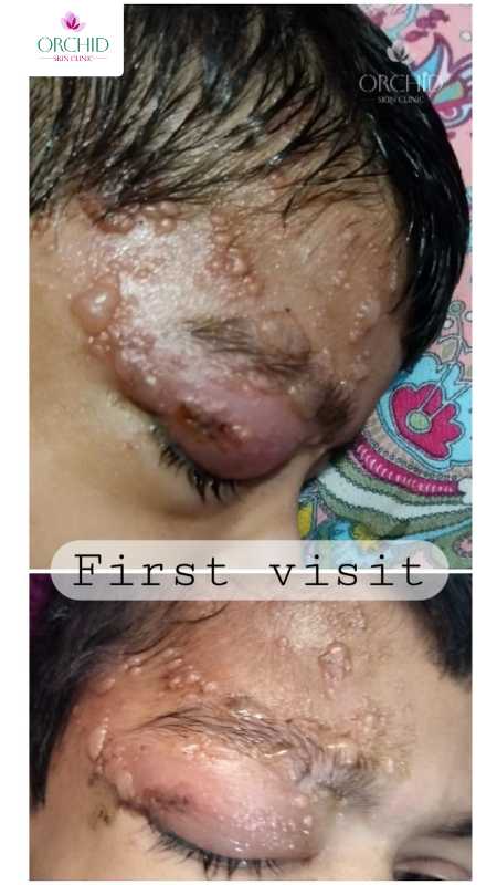
First Visit
Marked periorbital edema with grouped vesicles and crusting over forehead, eyebrow, and upper eyelid in V1 dermatome distribution. Significant facial swelling and periocular involvement evident.
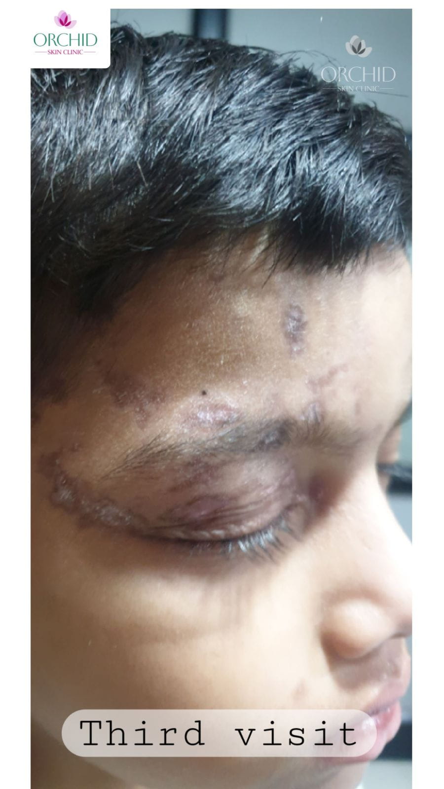
Third Visit (2-3 weeks)
Excellent response to early systemic antiviral therapy with marked resolution of edema, crust separation, and lesion crusting. Periorbital swelling significantly reduced demonstrating efficacy of valacyclovir therapy.
Clinical Significance of Serial Photography
Serial photography demonstrates the rapid therapeutic response to early-initiated valacyclovir therapy, preventing progression to sight-threatening complications such as keratitis or uveitis. The dermatomal restriction to V1 and the dramatic improvement in periocular inflammation underscore the importance of early recognition and treatment initiation.
Pathophysiology
Following primary varicella infection, VZV remains latent within dorsal root ganglia or cranial nerve ganglia. Reactivation in the trigeminal ophthalmic branch initiates a complex cascade:
- Neurogenic inflammation → Release of neuropeptides and cytokines
- Vesiculobullous rash → Characteristic grouped vesicles in dermatomal distribution
- Viral virulence → Risk of ocular tissue invasion and inflammation
- Immune response activation → T-cell mediated immunity
In pediatric cases, reactivation triggers include febrile illness, minor trauma, or residual immune immaturity following primary infection.
Treatment Approach
Systemic Antiviral Therapy
Primary Therapeutic Agent
Valacyclovir (weight-adjusted dosing)
- Initiated immediately upon diagnosis
- Treatment window: Critical within 72 hours of rash onset
- Significantly reduces ocular complications and post-herpetic neuralgia
- Dosing based on age and renal function
Supportive Care and Symptom Management
- Analgesics: Oral paracetamol and serratiopeptidase for pain relief and anti-inflammatory effects
- Local comfort: Cool compresses to affected areas
- Hydration: Adequate fluid intake and rest
- Topical therapy: Calamine lotion for lesion soothing and crust management
- Skin protection: Protective ointment to periocular skin to minimize secondary irritation
Ophthalmologic Monitoring
Serial ophthalmologic evaluations were performed at regular intervals to monitor for potential complications:
- Keratitis (viral keratitis or secondary bacterial)
- Anterior uveitis with or without secondary glaucoma
- Increased intraocular pressure
- Optic neuritis or posterior segment involvement
Additional Therapeutic Modalities
Reserved for complication management as needed:
Adjunctive Treatment Options
- Keratitis/Uveitis: Topical corticosteroids and cycloplegic agents under ophthalmologist supervision
- Severe neuropathic pain: Gabapentin or pregabalin if needed
- Immunocompromised patients: IV acyclovir for systemic protection
- Secondary bacterial infection: Topical or systemic antibiotics
- Post-herpetic neuralgia: Neuropathic pain modulators (rare in pediatric cases)
Clinical Course and Timeline
The patient demonstrated excellent response to early intervention with marked improvement by the third clinical visit (2-3 weeks post-treatment initiation):
Week 1-2: Early Intervention Phase
Initiation of valacyclovir and supportive care. Patient reported pain reduction and stabilization of lesion progression. No evidence of ocular involvement on examination.
Week 2-3: Regression Phase
Marked resolution of swelling and crusting. Lesions beginning to heal with minimal scarring. Pain substantially reduced. Continued ophthalmologic surveillance showed no complications.
Week 3+: Resolution Phase
Complete resolution of active vesicles with only minor post-inflammatory hyperpigmentation. Normal visual acuity maintained. No ocular sequelae. Excellent cosmetic outcome achieved.
The rapid clinical response highlights the critical importance of early antiviral intervention in preventing sight-threatening complications.
Clinical Significance and Key Takeaways
Important Clinical Implications
- Pediatric HZO rarity: Though uncommon in children, requires high index of suspicion with periocular vesicular rash
- Vision preservation: Early systemic antiviral therapy is the cornerstone of preventing corneal and retinal complications
- Multidisciplinary approach: Collaboration between dermatology and ophthalmology is mandatory for optimal outcomes
- Early recognition value: Education of patients and caregivers about early presentation in future events is critical
- Treatment window: Initiation within 72 hours of rash onset dramatically improves prognosis
- Monitoring importance: Regular ophthalmologic surveillance can detect subclinical complications
Prompt recognition and multidisciplinary management dramatically improved recovery and preserved this child's vision long-term.
Conclusion
This case of herpes zoster ophthalmicus in a 6-year-old child demonstrates the successful management of a potentially sight-threatening viral infection through early systemic antiviral therapy (valacyclovir), comprehensive supportive care, and strict ophthalmologic supervision. Early intervention resulted in rapid clinical resolution with complete healing, no ocular sequelae, preservation of normal visual acuity, and an excellent cosmetic outcome.
The key determinants of successful outcome were:
- Rapid clinical recognition and diagnosis
- Immediate initiation of appropriate antiviral therapy
- Multidisciplinary care coordination
- Close ophthalmologic follow-up
- Patient and caregiver education
Early antiviral therapy remains the critical determinant of prognosis in pediatric herpes zoster ophthalmicus. Delayed treatment significantly increases the risk of vision-threatening complications that may result in long-term visual morbidity.